The Relationship Between the QLs and Iliopsoas
63% of massage clients seek treatment for pain management which is a huge development in the industry, which was initially more thought of as a relaxation therapy. The iliopsoas and the QL are often the key to solving hip and back dysfunctions.
Most therapists will only really work the iliacus, and the lateral aspect of the QLs, rather than the iliopsoas and the more medial aspect of the QLs , but this approach will be very limited.
It will be more effective and less painful, to work firstly on the iliopsoas in its shortened state before proceeding to manipulation in an extended position. Then you can start to work the QLs laterally, working medially towards the spine.
THE ILIOPSOAS
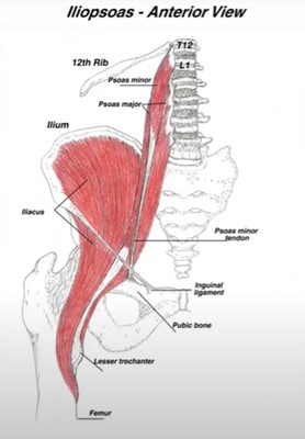
The iliopsoas, combining the iliacus and the psoas, attaches the upper (T12) and lower (femur) body, and the anterior and posterior, so it communicates in a wide reaching way.
THE PSOAS
Attachments
- lateral aspects of the T12 vertebrae and all lumbar evrtebrae and corresponding discs
- lesser trochanter of femur
Functions
- aids in anchoring the head of the femur in the acetabulum
- contains both slow and fast twitch fibres
Innervation
Lumbar plexus spinal nerves L2, L3 and L4
Psoas Minor
The psoas minor is absent bilaterally in 40- 50% of the population. The presence of a psoas minor can assist in childbirth.
Attachments
T12 to L2 vertebral body and corresponding discs to superior ramus of pubis
Innervation
Lumbar plexus spinal nerve L1
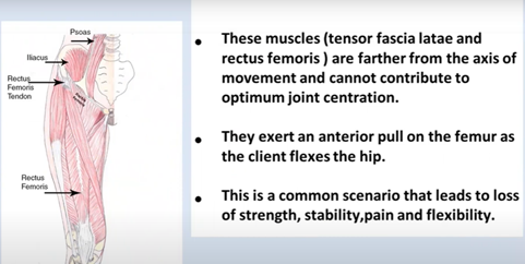
The psoas and psoas minor have similar attachments but the psoas minor does not cross the hip joint. Inhibition of the iliopsoas due to a torn labrum or disc irrittion can cause overuse of the tensor fascia latae and rectus femoris.
THE ILIACUS
Attachments
- upper 2/3 of the inner surface of the iliac fossa, anchoring to the lip of the iliac crest completely lining the wall of the pelvis
- anterior portions of the sacroiliac and iliolumbar ligaments
- the iliacus fibres connect with the psoas to attach to the lesser trochanter and the capsule of the hip joint
Innervation
Lumbar plexus spinal nerves L2 and L3
Common actions of the iliopsoas include:-
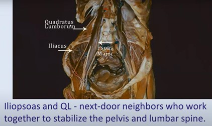
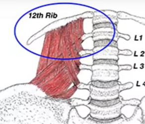
There is a close relationship between the iliacus, the QL and the psoas. The QL attaches at the 12th rib and psoas at T12.The QL attaches to the posterior lip of the ilium and the iliacus to the anterior. They work together to stabilise the pelvis and the lumbar spine.
Also the same nerve that innervates the psoas, also innervates the kidneys, so work here may help to calm the central nervous system where there has been kidney stones, lower back pain, testicular or erectile dysfunction.
The psoas is also one of the earliest muscles to form in utero – it brings upper and lower body together; the foetus is in a flexed position.
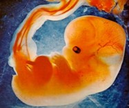
Because of its early development and the shape that it puts us in, it is also connected to the fight, flight or freeze response, so we must acknowledge a sense of client safety when working on this muscle.
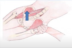
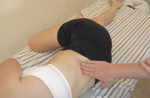
TECHNIQUES
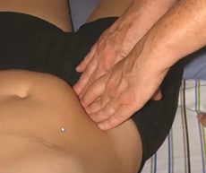
Take a lateral approach and slowly work more medially, rather than working anteriorly to posteriorly through the rectus abdominis. Taking a slow lateral to medial approach will be less painful and more effective, while maintaining client safety.
Why does the iliopsoas sometimes contribute to neck pain?
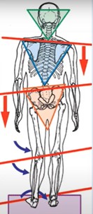
- unilateral psoas spasm causes contralateral rotation and ipsilateral side bend
- in order to heal the pulled neck muscles, the twist in the core has to be corrected first
THE QUADRATUS LUMBORUM
The QLs don’t only cause back and pelvic pain, but if shortened can also cause hip hike, poor abduction and a low shoulder.
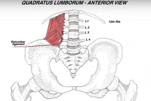
Attachments
- 12th rib and L1 -L4
- ilium and iliolumbar ligament
Innervation
Ventral rami of the T12 and L1, L2, L3 and L4 spinal nerves
Functions
- lateral flexion of the spine
- elevation of the ipsilateral ilium (hip hiking)
- contributes to extension of the spine
- stabilizes the 12th rib during inhalation and forced exhalation
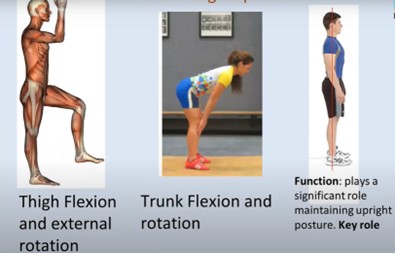
- plays a significant role in maintaining upright posture
The QL is a stabilising muscle which has a vital role in maintaining an upright posture.

The QL has a complex fibre arrangement, making the muscle much tougher so that it can act as a stabiliser. It stabilises the 12th rib during inhalation and exhalation, hence why sometimes someone may cough with a lot of force and then not be able to stand up straight.
The QLs, through satellite gluteus medius trigger points, are often responsible for pseudo disc syndrome and failed surgical back syndrome.
As infants we go through developmental stages of movement eg. pushing and reaching. The push must precede the reach or the reach results in falling over or collapsing. The QL is a push muscle, ie. stabilizes, brings strength and supports us internally and externally.
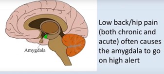
The QL, like the iliopsoas, also bridges the upper and lower body, stabilising the hip and lower spine. when there is pain in these areas, the amygdala goes on high alert, sensing danger and initiating the fight or flight response, so again we must maintain emotional safety for the client.
If a QL is overactivated, this can cause a weak and inhibited gluteus medius on the opposite side, which may even cause instability in the ankle joint.
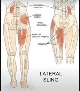
The lateral sling stabilises the frontal plane, hence why overactivation on one side can cause issues on the opposite side.
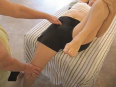
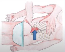
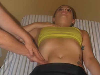
TECHNIQUES
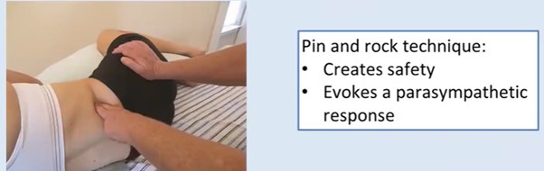
Pin the muscle with the fingers and then rock the hip, and as the relaxation response kicks in, the muscle should start to relax.
The running man technique to work the medial QL fibres. Gently pin the muscle with the fingers, and ask the client to gently flex and extend the hip.
SUMMARY OF THE QL AND ILIOPSOAS
- The iliopsoas and QL have extensive bodywide effects
- Gentle, slow techniques can be more effective
- Both muscle have deep body/mind considerations ie require emotional safety